
SOMATOM® On.site
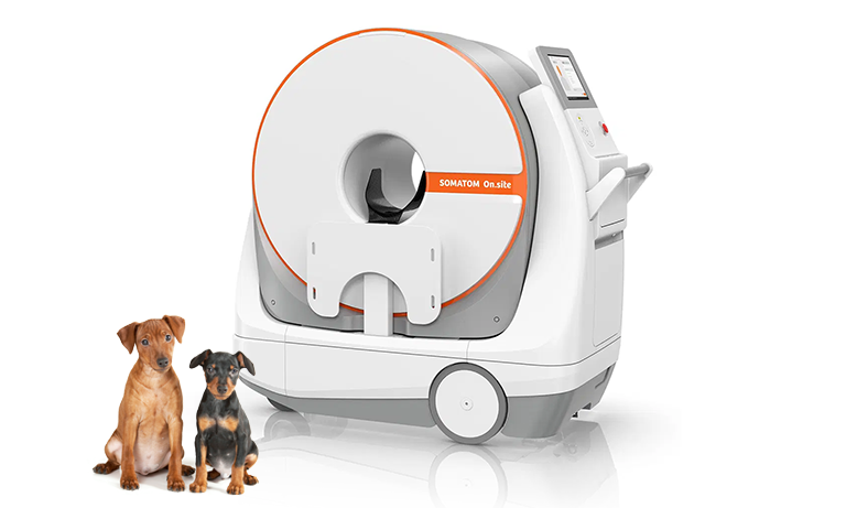
Bringing critical care imaging to your patient with a portable Veterinary CT
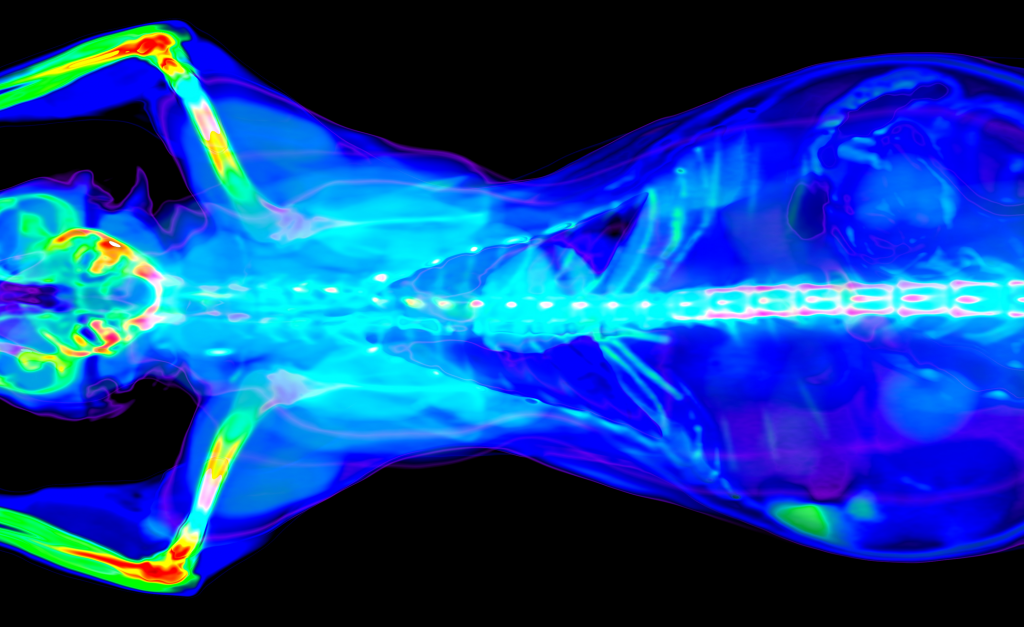
The Siemens SOMATOM On.site revolutionizes veterinary diagnostic systems by delivering high-quality imaging directly to your practice. With its easy scan setup, fast workflow, and integrated patient support accessories, it ensures convenient and consistent patient positioning. The integrated drive camera offers real-time viewing on the built-in Touch UI display, simplifying maneuverability for the technologist.
The scanner’s distinctive telescopic gantry design moves the radiation source away from the patient during scanning, unlike conventional mobile CT scanners that do not utilize such a design concept. This advanced technology enhances safety without compromising performance.
At Universal Systems Diagnostics, Inc, we offer both new and refurbished systems, meeting a variety of large and small animal veterinary imaging needs and budgetary considerations.
Key Features:
- All-in-one System with Integrated computers for further flexibility for patient scans in small spaces where additional equipment would be a hindrance. The slim system with built-in trolley & batteries includes integrated patient support accessories
- Smart driving concept with motorized trolley with an ergonomic drive handle allows easy driving and maneuvering
- Ample space for patient positioning
- Easy Scan set-up and fast workflow
– Intuitive Touch UI
– GO technologies provide guidance for cross-trained staff through the scanning process
– Recon&GO performs automated postprocessing and automatically uploads the images to PACS
-Single-operator design - Telescopic scanner that glides on the stationary system trolley base during the scanning process.
- Self-shielded system including attachable radiation shields to cover the front and back gantry openings
Specifications:
- Detector: 2.4 cm (Stellar UFC)
- Physical detector rows: 32
- Detector row thickness: 0.75 mm
- Acquisition mode: Spiral
- Tube voltage: 80, 120 kV
- Tube current: 3-25 mA (increments of 1 mA)
- kW output/generator power: 3 kW
- Gantry aperture: 35 cm
- Scan field/FOV: 26 cm, HD FOV 35 cm*
- Scan range: 22.3 cm
- Pitch: 0.35 – 1.5 (in steps of 0.05)
- Iterative reconstruction algorithms: ADMIRE, iMAR
- Reconstruction slice thickness options: 0.8, 1.0, 1.5, 2.0, 3.0, 4.0, 5.0, 6.0, 7.0, 8.0, 10.0 mm
- Tube cooling: Hybrid (air & water)
- Image transfer: Wireless and LAN connection to hospital network
- Batteries:1 rechargeable lithium-ion battery (1 x 48 V)
*The image quality for the area outside the 26 cm scan field of view does not meet the image quality of the area inside the 26 cm scan field of view. Image artifacts may appear, depending on the patient setup and anatomy scanned. HD FOV cannot be used for scan FOV smaller than 26 cm.
SIEMENS VETERINARY CT
SOMATOM On.site
The Siemens SOMATOM On.site revolutionizes veterinary diagnostic systems by delivering high-quality CT imaging directly to your practice. Its telescopic gantry design, easy scan setup, and fast workflow make it the world’s first truly portable veterinary CT — bringing critical care imaging to the patient instead of the other way around.
MOBILE CT, NO ROOM REQUIRED, 32-SLICE, TELESCOPIC GANTRY, SINGLE-OPERATOR, BATTERY POWERED
DETECTOR ROWS
32 rows
.75 mm thickness
GANTRY APERTURE
35 cm
Telescopic design
SCAN FIELD (FOV)
26 cm
HD FOV up to 35 cm
GENERATOR POWER
3 kW
3-25 mA output
POWER SOURCE
Battery
48V lithium-ion
OVERVIEW
CT imaging that comes to the patient
The Siemens SOMATOM On.site revolutionizes veterinary diagnostic systems by delivering high-quality imaging directly to your practice. With its easy scan setup, fast workflow, and integrated patient support accessories, it ensures convenient and consistent patient positioning. The integrated drive camera offers real-time viewing on the built-in Touch UI display, simplifying maneuverability for the technologist.
The scanner’s distinctive telescopic gantry design moves the radiation source away from the patient during scanning — unlike conventional mobile CT scanners that don’t use this design concept. This advanced technology enhances safety without compromising performance, and is available from USD in both new and remanufactured configurations.
Truly portable CT
All-in-one system with built-in trolley and batteries — no dedicated CT room or specialized power infrastructure required.
Telescopic gantry safety
The radiation source moves away from the patient during scanning — a design advantage over conventional mobile CT systems.
Single-operator workflow
GO technologies guide cross-trained staff through scanning, while Recon&GO automates postprocessing and PACS upload.
Wireless image transfer
Wireless and LAN connectivity to your hospital network means images move as easily as the scanner itself.
TECHNICAL SPECIFICATIONS
System Specifications
Detector
Detector
2.4 cm (Stellar UFC)
Physical detector rows
32
Detector row thickness
.74 mm
Gantry aperture
35 cm
Scan field / FOV
26 cm, HD FOV 35 cm*
Scan range
22.3 cm
X-Ray System
Acquisition mode
PUREINSIGHT
Tube voltage
320 rows
Tube current
.5 mm
Generator power
16 cm
Pitch
1024
Tube cooling
1024
Imaging
X-Ray tube
CoolNovus
Generator power
100 kW
Maximum mA output
Up to 1,400 mA
kV selection
70, 80, 100, 120, 135 kV
Dose reduction
SilverBeam filter (Ultra-low dose)
Reconstruction
AiCE Whole Body Deep Learning
Mobility & Power
Couch weight capacity
315 kg / 695 lbs
Tabel motion
Motorized, bidirectional
Lateral slide
17 cm total (Tech Assist)
Compatible patients
Small to large animals
CLINICAL APPLICATIONS
What this system is built for
The Aquilion ONE / INSIGHT Edition is suited for a wide range of veterinary clinical applications across companion animals and larger patients.
Cardiac imaging
Fast rotation and wide coverage enable complete cardiac studies in a single breath-hold equivalent scan cycle.
Neurological imaging
High-resolution brain and spinal cord imaging with PIQE 1024 matrix for detailed soft tissue differentiation.
Oncology studies
Whole-body coverage in a single pass supports comprehensive oncological staging and treatment planning.
Emergency & trauma
Sub-second scan times enable rapid whole-body trauma surveys for critically ill or injured patients.
RELATED SYSTEMS
Other Siemens CT systems
SIEMENS
SOMATOM go.Up
Advanced spectral CT with AI-powered reconstruction for superior material differentiation.
SIEMENS
SOMATOM go.All
Advanced spectral CT with AI-powered reconstruction for superior material differentiation.
SIEMENS
SOMATOM go.Top
Whole-organ CT with advanced detector technology and dynamic scanning capability.
SIEMENS
SOMATOM go.Now
Ultra-high resolution CT with submillimeter detail for small animal neurological imaging.