

EqueTom™
Tomosynthesis imaging technology for the equine market.
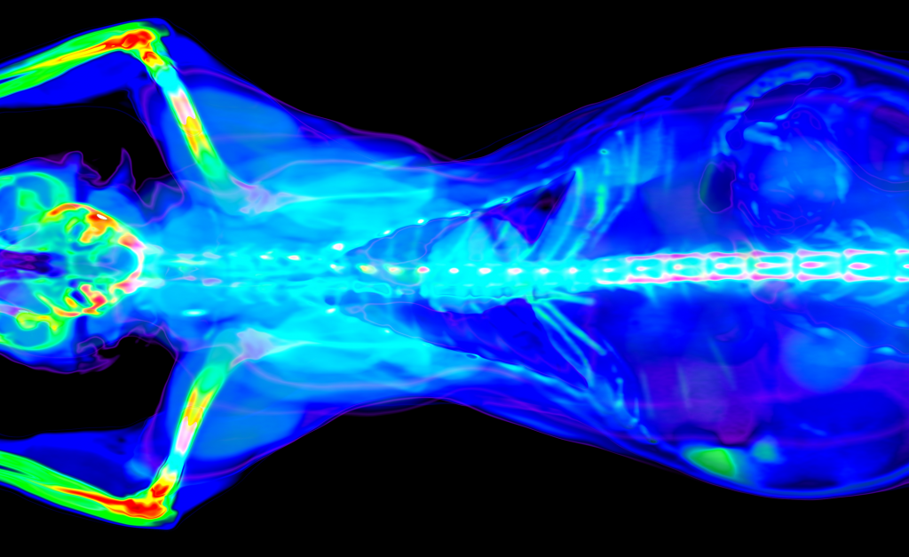
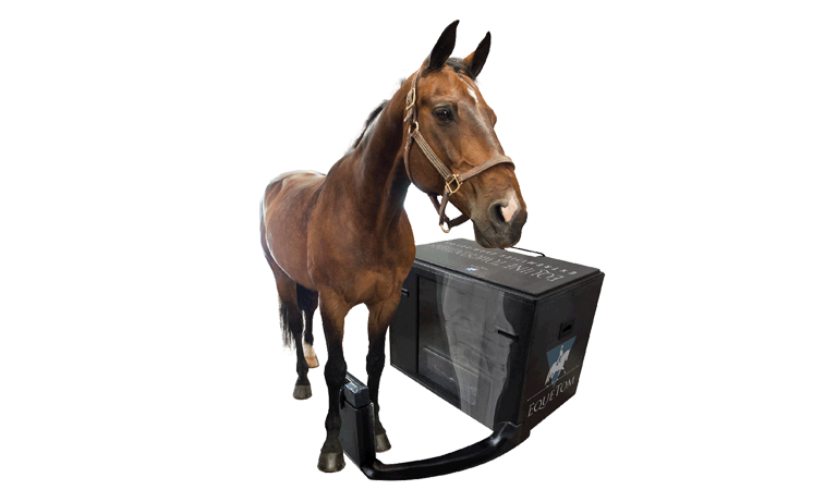
The Orimtech EqueTom™ is a system using the tomosynthesis imaging technology developed for the equine market. It offers high-quality low-dose multi-slice images to visualize the parts of the objects that are invisible by the conventional 2D radiography

Key Features:
- The unique reconstruction technique provides about 240 coronal slices of the examined body part for detailed examination, instead of a single static X-Ray.
- Designed to be portable. It can be carried between stables and work the same routine as with digital radiography systems with in addition the ability to acquire tomosynthesis slices of horse leg.
- Battery powered. Battery life up to 50 tomosynthesis exams and 100 radiography shots.
- For the body-parts where tomosynthesis is not applicable the portable generator and digital detector can be removed and take radiography shots.
Specifications:
- Below 3 seconds images acquisition technique in tomosynthesis mode and ~1-minute 3D reconstruction time provide fast and accurate diagnostic information. Focus at different depth inside the body via swiping the slices using our user-friendly graphic interface on your laptop.
- Scanable anatomy (tomosynthesis): Heel, Pastern, Ergot, Cannon, Carpus, Hoof, Coronet, Fetlock, Hock, Teeth, Sinuses, TMJ
- Scanable anatomy (radiography): all body-parts.
- Radiography resolution 125 μm
- Generator type: battery powered high-frequency 1.6 kW.
- Acquisition frame rate of up to 35 frames per second and tomosynthesis angles about ±15°.
- Spatial resolution within slice: 300 μm.
- Slice thickness (pitch): 0.5-8 mm
ORIMTECH VETERINARY CT
EqueTom™
The Orimtech EqueTom™ is a portable, battery-powered X-ray tomosynthesis system purpose-built for equine applications, bringing multi-slice 3D imaging to the stable without the infrastructure, cost, or complexity of a conventional CT installation. Using Orimtech’s proprietary ACE/AVRG reconstruction technology, it produces approximately 240 coronal slices of any equine distal limb anatomy in under 3 seconds of acquisition time, delivering diagnostic detail that conventional 2D radiography simply cannot provide. With up to 50 tomosynthesis exams and 100 radiography shots per battery charge, it works exactly like a digital radiography system, with the added power to see inside the structures that matter most.
TOMOSYNTHESIS, PORTABLE, BATTERY-POWERED, 240 SLICES, 300 µm RESOLUTION, EQUINE DISTAL LIMB, DICOM COMPATIBLE
IMAGING MODE
Tomosynthesis
SLICES
240
ACQUISITION TIME
<3 sec
SPATIAL RESOLUTION
300 µm
BATTERY CAPACITY
50 / 100
TECHNICAL SPECIFICATIONS
System Specifications
Imaging
Imaging modalities
Tomosynthesis + digital radiography
Tomosynthesis slices
240 coronal slices per acquisition
Acquisition time
Under 3 seconds (tomosynthesis mode)
3D reconstruction time
1 minute
Spatial resolution (in-slice)
300 µm
Radiography resolution
125 µm
Slice thickness (pitch)
0.5–8 mm
Tomosynthesis scan angle
±15°
Frame rate
Up to 35 frames per second
Reconstruction engine
Orimtech ACE/AVRG proprietary technology
DICOM / PACS
Full compatibility
X-Ray System
Generator type
Battery-powered high-frequency generator
Generator power
1.6 kW
Power source
Battery, no external power supply required
Detector
Digital flat-panel detector (removable)
Scannable Anatomy
Tomosynthesis regions
Heel, pastern, ergot, cannon, carpus, hoof, coronet, fetlock, hock, teeth, sinuses, TMJ
Radiography regions
All body parts, generator, and detector detach for standard DR
Species
Equine, purpose-built for horses
Portability
Portability
Fully portable, designed to move between stables
Battery capacity (tomo)
Up to 50 tomosynthesis exams per charge
Battery capacity (DR)
Up to 100 radiography shots per charge
Workflow
Same routine as digital radiography, with added tomosynthesis capability
User interface
Laptop-based, swipe to focus at different depths inside the body
CLINICAL APPLICATIONS
What this system is built for
The EqueTom is built for equine veterinary professionals who need multi-slice 3D imaging at the point of care, delivering tomosynthesis detail of the distal limb in a portable, battery-powered system that travels between stables as easily as a standard digital radiography unit.
Distal limb tomosynthesis
Approximately 240 coronal slices of the equine distal limb are acquired in under 3 seconds, revealing heel, pastern, fetlock, hoof, coronet, carpus, hock, and cannon anatomy at 300 µm in-slice spatial resolution that exposes pathology invisible to conventional 2D radiography.
Hoof & coffin joint imaging
Tomosynthesis slices of the hoof, coronet, and coffin joint region allow veterinarians to focus on different depths within the tissue by swiping through slice planes on a laptop, resolving fine cortical detail, subchondral bone changes, and soft tissue structures that a single static X-ray collapses into a single overlapping projection.
Teeth, sinuses & TMJ
Beyond limb anatomy, the EqueTom supports tomosynthesis acquisition of equine teeth, paranasal sinuses, and the temporomandibular joint, extending its diagnostic utility to dental and head imaging applications that benefit from depth-selective slice viewing without the need for anesthesia or a fixed imaging facility.
Portable field radiography
When tomosynthesis is not required, the generator and digital detector detach from the system for use as a standalone digital radiography unit, covering all equine body parts and maintaining 125 µm radiographic resolution. One battery charge supports up to 100 radiography shots, making the EqueTom a complete portable imaging solution for equine field practice.
RELATED SYSTEMS
Other Orimtech CT systems
ORIMTECH
EDAMIS
Advanced spectral CT with AI-powered reconstruction for superior material differentiation.
ORIMTECH
MAISEES
Advanced spectral CT with AI-powered reconstruction for superior material differentiation.