
ORIMTECH VETERINARY CT
MAISEES
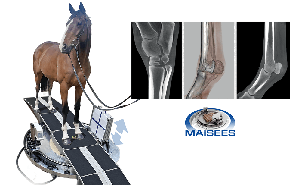
The Orimtech MAISEES is a multimodality adaptive imaging system for equine extremity studies, combining cone beam CT, digital radiography, and fluoroscopy in a single integrated platform that adapts its field of view to the desired anatomy through source and detector motion modulation, without requiring the horse to move or be repositioned. With 300 µm isotropic 3D spatial resolution, a 15-second expedited CT acquisition mode, and built-in motion registration and compensation that enables diagnostic-quality imaging with only light sedation, the MAISEES redefines what is possible in standing equine CT, making high-quality three-dimensional extremity imaging accessible to equine facilities without the infrastructure, anesthesia risk, or operational complexity of conventional fixed-gantry equine CT systems.
CBCT, ADAPTIVE FOV, STANDING EQUINE, 300 MICRON RESOLUTION, 15-SECOND SCAN, RADIOGRAPHY, FLUOROSCOPY
CT FOV
D270 mm
SPATIAL RESOLUTION
300 micron
SCAN TIME (FAST)
15 sec
SCAN TIME (HQ)
30 sec
KV RANGE
40–120 kV
TECHNICAL SPECIFICATIONS
System Specifications
CT Specifications
CT field of view
D270 mm × H270 mm
Isotropic 3D spatial resolution
300 micron
Scan time (expedited mode)
15 seconds
Scan time (high quality mode)
30 seconds
X-ray technique
40–120 kV / 0.1–0.3 mAs per frame
DICOM / PACS
Full compatibility, patient database included
Adaptive FOV
FOV adaptation
Source and detector motion modulation adapts FOV to desired extremity location
Animal movement required
None, FOV adapts to anatomy without repositioning the horse
Anatomy access
Any distal limb location, the system moves to the target, not the patient
Motion compensation
Integrated motion registration and compensation for high spatial resolution despite patient movement
Workflow
Sedation requirement
Light sedatives only, no deep anesthesia required
Animal positioning
Natural standing position, no repositioning of the horse needed
FOV targeting
Automated source and detector motion adapts to desired anatomy
User interface
Full DICOM patient database with PACS compatibility
All Modalities
CT (CBCT)
300 micron isotropic 3D, 15 or 30 second acquisition
Radiography
Digital radiography of equine extremities
Fluoroscopy
Real-time fluoroscopic imaging of equine extremities
Modality switching
Single integrated system, all modalities available without reconfiguration
CLINICAL APPLICATIONS
What this system is built for
The MAISEES is designed for equine veterinary facilities that need fast, high-quality 3D extremity imaging with only light sedation, delivering adaptive cone beam CT, radiography, and fluoroscopy of any distal limb location without requiring the horse to be repositioned between acquisitions.
Adaptive standing equine CT
The system’s source and detector motion modulation automatically adapts the field of view to the desired extremity location, without moving or repositioning the horse. Any distal limb anatomy from the carpus and tarsus can be targeted in sequence during a single standing sedation session, reducing stress on the patient and total examination time for the clinical team.
Rapid CT acquisition for difficult patients
A 15-second expedited scan mode and built-in motion registration and compensation allow diagnostic-quality 300-micron isotropic CT to be acquired even when the horse shifts weight or moves during scanning, making the MAISEES practical for patients that would be difficult or unsafe to hold still for longer acquisition protocols, without resorting to deep anesthesia.
Lameness & orthopedic diagnosis
300-micron isotropic 3D spatial resolution across a D270 mm × 270 mm field of view resolves fine bone and soft tissue detail of the equine distal limb, enabling confident diagnosis of subchondral bone disease, collateral ligament injury, coffin joint pathology, navicular syndrome, and fetlock arthropathy in a single integrated imaging session combining CT, radiography, and fluoroscopy.
Real-time fluoroscopy & radiography
Integrated real-time fluoroscopy supports dynamic joint assessment, contrast studies, and guided injection procedures during the same examination session as CT, while digital radiography provides high-resolution 2D images for direct comparison with tomographic findings. The combination of all three modalities in a single system eliminates the need to transport the horse between imaging suites for different study types.
RELATED SYSTEMS
Other Orimtech CT systems
ORIMTECH
EqueTom
Advanced spectral CT with AI-powered reconstruction for superior material differentiation.
ORIMTECH
EDAMIS
Advanced spectral CT with AI-powered reconstruction for superior material differentiation.