
ORIMTECH VETERINARY CT
EDAMIS
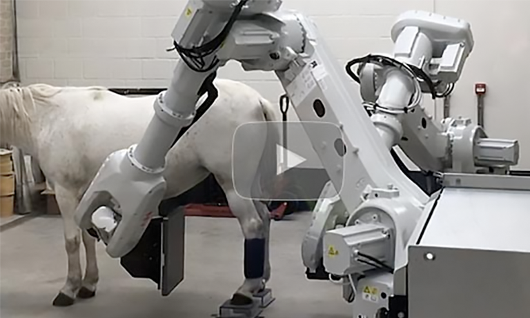
The Orimtech EDAMIS is a dual-arm robotic multi-modality imaging system for equine applications integrating cone beam CT, fluoroscopy, tomosynthesis, and digital radiography into a single platform built on Orimtech’s ACE/AVRG technology. Designed to scan horses in a natural standing position with only light sedation, it eliminates the anesthesia risk of conventional equine CT while delivering 250-micron isotropic CBCT resolution, ±25 HU accuracy, and the full flexibility of a robotically controlled source-detector system that can adapt its orientation and scanning trajectory to any anatomy. For equine hospitals and specialty referral facilities requiring the full spectrum of imaging modalities in one integrated system, nothing else compares.
CBCT, FLUOROSCOPY, TOMOSYNTHESIS, RADIOGRAPHY, STANDING EQUINE, 250 MICRON RESOLUTION, NO GENERAL ANESTHESIA, DUAL-ARM ROBOTIC
MODALITIES
4-in-1
CBCT SPATIAL RESOLUTION
250 micron
CBCT FOV
D240 mm x 240 mm
SCAN ANGLE
210°
HU ACCURACY
±25 HU
TECHNICAL SPECIFICATIONS
System Specifications
CBCT
Spatial resolution
250 micron (mostly defined by a tube focal spot)
Scan angle
210°
Field of view (single slab)
D240 mm × 240 mm
Field of view (stitched)
D240 mm × 420 mm (2 stitched slabs, optional)
Single slab scan time
10–60 seconds depending on X-ray dosage
Acquisition technique
Pulsed — up to 25 FPS
Scanning trajectories
Circular and non-circular
HU accuracy
±25 HU on CTP486 section (CATPHAN)
Motion artifact mitigation
Integrated Qualysis visual motion tracking subsystem
Reconstruction engine
AVRG with support of scatter, beam-hardening, and metal artifact reduction
3D image viewer
Orimtech MPR proprietary; Fovia HDVR optional
Tomosynthesis
Spatial resolution
200 micron (mostly defined by a tube focal spot)
Scan angle
15–90° (angular); 360° (circular motion)
Field of view
D240 mm × 240 mm
Scan time
4–20 seconds
Acquisition technique
Pulsed, up to 25 FPS
Scanning trajectories
Circular and non-circular
Motion artifact mitigation
Integrated Qualysis visual motion tracking subsystem
Reconstruction engine
AVRG (Orimtech)
3D image viewer
Orimtech proprietary
Fluoro
Automatic brightness control
Hardware and software supported
Real-time display
Supported with Orimtech proprietary dynamic contrast enhancement
Playback
Supported with Orimtech proprietary dynamic contrast enhancement
FOV position control
Joystick, PC keyboard, and touch screen
Subtractive fluoroscopy
Supported for contrast studies
Radiography
Spatial resolution
~5 LP/mm (XRD 4343 RF)
Object positioning
Arbitrary within the system mechanical accessibility
Source-detector distance
Arbitrary within the system mechanical accessibility
FOV adjustment
Collimator light and laser; joystick-controlled system motion
DR image processing
Orimtech proprietary library; CVIE (Context Vision) ready
Common Features
System design
Dual-arm robotic, full flexibility in source-detector orientation and trajectory
Equine application
Standing position scanning — only light sedation required
Anatomy (extremities)
Natural standing position — built-in motion capture and compensation
Anatomy (head/neck)
400 micron isotropic resolution for dental applications
Dose modulation
X-ray technique modulation during scan for optimal dose utilization
Collimation
4-blade PC-controlled automatic collimator
DICOM support
PACS compatible
Supported generators
Swiss Genesis (Betschart), CPI Indico IQ 100, COMET
Supported detectors
Varex XRD 4343 RF; iRay Mercu1717V
CLINICAL APPLICATIONS
What this system is built for
The EDAMIS is purpose-built for equine hospitals and referral facilities that need the full range of diagnostic imaging modalities, cone beam CT, fluoroscopy, tomosynthesis, and radiography, in a single integrated platform that scans horses in a standing position without the risk and cost of general anesthesia.
Standing equine CBCT:
250-micron isotropic cone-beam CT of equine extremities and head is acquired in 10–60 seconds with the horse in a natural standing position, requiring only light sedation. Built-in Qualysis motion tracking and compensation ensure high spatial resolution images even when the patient moves, eliminating the primary limitation of standing equine CT.
Equine head, dental & neck CT
The dual-arm robotic design adapts its orientation to image the equine head and neck, in addition to extremities, delivering 400 micron isotropic resolution for dental applications with X-ray technique modulation for optimal penetration of dense dental and craniofacial structures. Sinus, dental arcade, and nasal passage pathology are evaluated without general anesthesia.
Real-time fluoroscopy & dynamic studies
Integrated fluoroscopy with Orimtech’s proprietary dynamic contrast enhancement enables real-time visualization of joint dynamics, contrast media flow, and functional anatomy, supporting tenoscopy guidance, contrast studies, and dynamic lameness evaluation in a single imaging session alongside CT and radiography.
Multi-modality workflow efficiency
With up to 12 user-predefined CBCT scanning trajectory protocols and 8 tomosynthesis protocols, full DICOM/PACS compatibility, and a single integrated acquisition system covering all four modalities, the EDAMIS eliminates the need for multiple specialized imaging systems, reducing capital investment, floor space requirements, and per-exam workflow complexity across the full spectrum of equine diagnostic imaging.
RELATED SYSTEMS
Other Orimtech CT systems
ORIMTECH
MAISEES
Advanced spectral CT with AI-powered reconstruction for superior material differentiation.
ORIMTECH
EqueTom
Advanced spectral CT with AI-powered reconstruction for superior material differentiation.